Original Dataset of Pulse Waves for Thousands of Virtual Subjects
The full database - together with Matlab functions for processing and analysing the data - can be downloaded from the links provided at the bottom of this page.
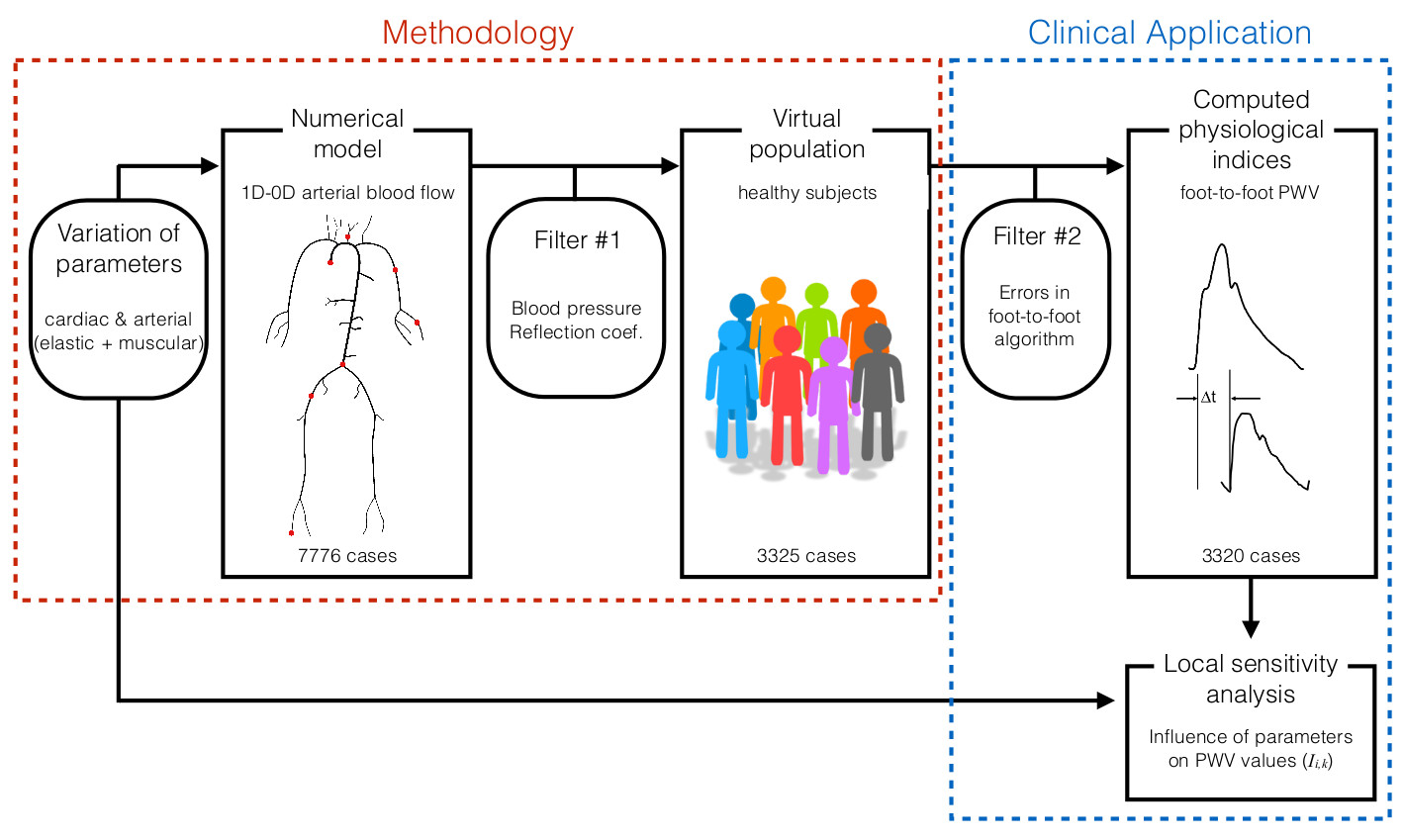
Using Nektar1D, we created a database of 3,325 virtual subjects, each with distinctive arterial pulse waveforms.
For each subject, blood pressure, blood flow and luminal area waveforms are available at multiple arterial locations, together with the parameters of the simulation (e.g. vessel geometries, cardiac output, pulse wave velocities).
 In our initial study (Am J Phys, 2015), we used the database to assess the accuracy of pulse wave indices of aortic pulse wave velocity (PWV). This is a surrogate for aortic stiffness which is an important marker of vascular health, as well as an important physical parameter for calculating central (aortic) blood pressure from non-invasive clinical data.
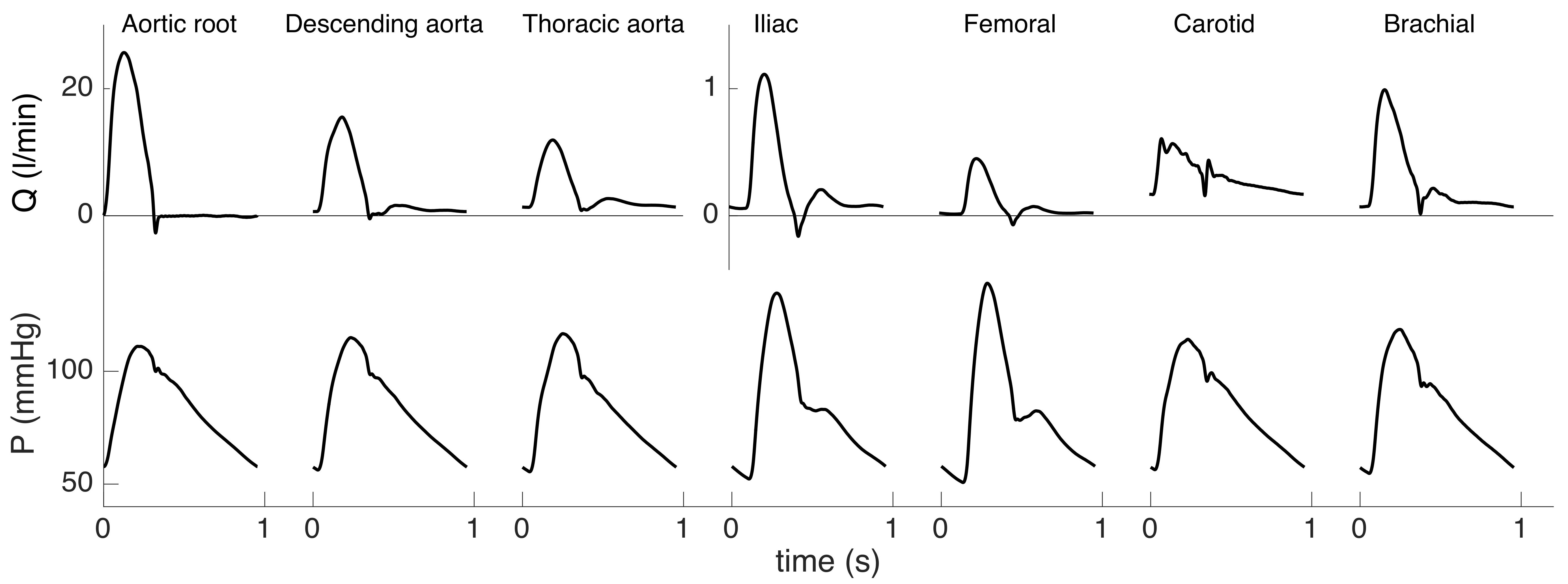
The following figure presents an example of simultaneous pressure and flow waves at several locations in the arterial network for one virtual subject. The main waveform features observed in vivo are reproduced, including amplification of the pulse pressure distal to the heart, positive carotid flow, and presence of backflow in early diastole in the leg arteries. The aortic pressure wave presents an inflection point located before the systolic peak; waveforms are therefore of type-A according to Murgo's classification, which are representative of an older age category.

A database of virtual healthy subjects to assess the accuracy of foot-to-foot pulse wave velocities for estimation of aortic stiffness.
American Journal of Physiology: Heart and Circulatory Physiology,
309:H663-675, 2015

While central (carotid-femoral) foot-to-foot pulse wave velocity (PWV) is considered to be the gold standard for the estimation of aortic arterial stiffness, peripheral foot-to-foot PWV (brachial-ankle, femoral-ankle, and carotid-radial) are being studied as substitutes of this central measurement. We present a novel methodology to assess theoretically these computed indexes and the hemodynamics mechanisms relating them. We created a database of 3,325 virtual healthy adult subjects using a validated one-dimensional model of the arterial hemodynamics, with cardiac and arterial parameters varied within physiological healthy ranges. For each virtual subject, foot-to-foot PWV was computed from numerical pressure waveforms at the same locations where clinical measurements are commonly taken. Our numerical results confirm clinical observations: 1) carotid-femoral PWV is a good indicator of aortic stiffness and correlates well with aortic PWV; 2) brachial-ankle PWV overestimates aortic PWV and is related to the stiffness and geometry of both elastic and muscular arteries; and 3) muscular PWV (carotid-radial, femoral-ankle) does not capture the stiffening of the aorta and should therefore not be used as a surrogate for aortic stiffness. In addition, our analysis highlights that the foot-to-foot PWV algorithm is sensitive to the presence of reflected waves in late diastole, which introduce errors in the PWV estimates. In this study, we have created a database of virtual healthy subjects, which can be used to assess theoretically the efficiency of physiological indexes based on pulse wave analysis.

In subsequent studies, the dataset has been used to produce algorithms for non-invasive estimation of
(i) local arterial wave speed and mean blood velocity waveforms
using Doppler ultrasound (Physiol Measur, 2017);
and
(ii) central blood pressure from the aortic flow (Hypertension, 2017; J Biomech, 2016).
Forward and backward pressure waveform morphology in hypertension has also been studied using this virtual subjects (Hypertension, 2017).
The algorithms used in all these studies were first developed and tested using virtual subjects and then assessed in vivo using cohorts of real subjects.
Download the database
The complete database can be downloaded using the links below. Data is sorted by arterial location and saved in Matlab formatted files of about 300 MB each (under the 'Physiological data' column). The structure of the database is explained in detail in the reference manual. Subjects with pulse waveforms that did not satisfy the filter criteria for physiological values of blood pressure and reflection coefficients are also provided for completeness (under the 'Non-Physiological data' column). The Fictive_database.mat file contains a description of the arterial network geometry, and values of computed haemodynamic indices (e.g. cardiac output, pulse wave velocity, pulse pressure). Search tools (Matlab functions) are also provided, which return subjects from the database with specific parameters and computed indices.


